

Comparative Neurological and Sensory Physiology
Manatee Sensory Sytems Research in Dr. Roger Reep’s Laboratory
Former Graduate Students: Kari Clifton, Iske Larkin, Chris Marshall, Alex Costidis, Joseph Gaspard
Collaborators:
- Gordon Bauer, New College of Florida
- David Mann, University of South Florida
Understanding the sensory capacities of manatees is important for two main reasons. First, it is essential if we are to effectively devise ways to avoid destructive human-manatee interactions. By gaining insight into manatee sensory capacities we can design warning devices that are tuned to those capacities. Of specific interest here is the system of tactile hairs (vibrissae) on the body, which may function as a mammalian lateral line to detect water movements and low frequency vibrations associated with boats, other animals, and water currents. This system is also hypothesized to aid navigation by helping to detect landmarks in the environment. A second reason to investigate manatee sensory systems is their importance in a comparative evolutionary context. Manatees (and sirenians generally) represent a novel branch of mammalian evolution, and the brains and behaviors of these animals likewise exhibit unusual and often unique traits. Thus, by elucidating the patterns of brain organization subserving these capacities we gain direct insight into the range of evolutionary potential in the mammalian lineage.
The current goals of this project are to investigate the role of vibrissae in Florida manatee perception and behavior, and in the organization of the nervous system.

A. Side view of cranial body; small arrowheads indicate hair follicle papillae.
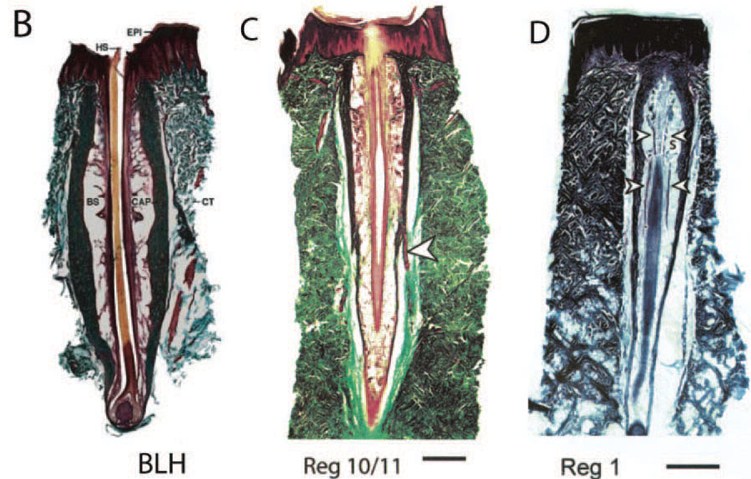
B. Longitudinal section of facial bristle-like hair follicle. BS=blood sinus, CAP=capsule, CT=connective tissue, EPI=epidermis, HS=hair shaft.
C. Postfacial follicle from ventral body, with extended cavernous sinus. Arrowhead indicates blood vessel. Scale bar=1mm.
D. Postfacial hair from dorsal body. Arrowheads indicate nerves, S is a blood sinus. Scale bar=1mm
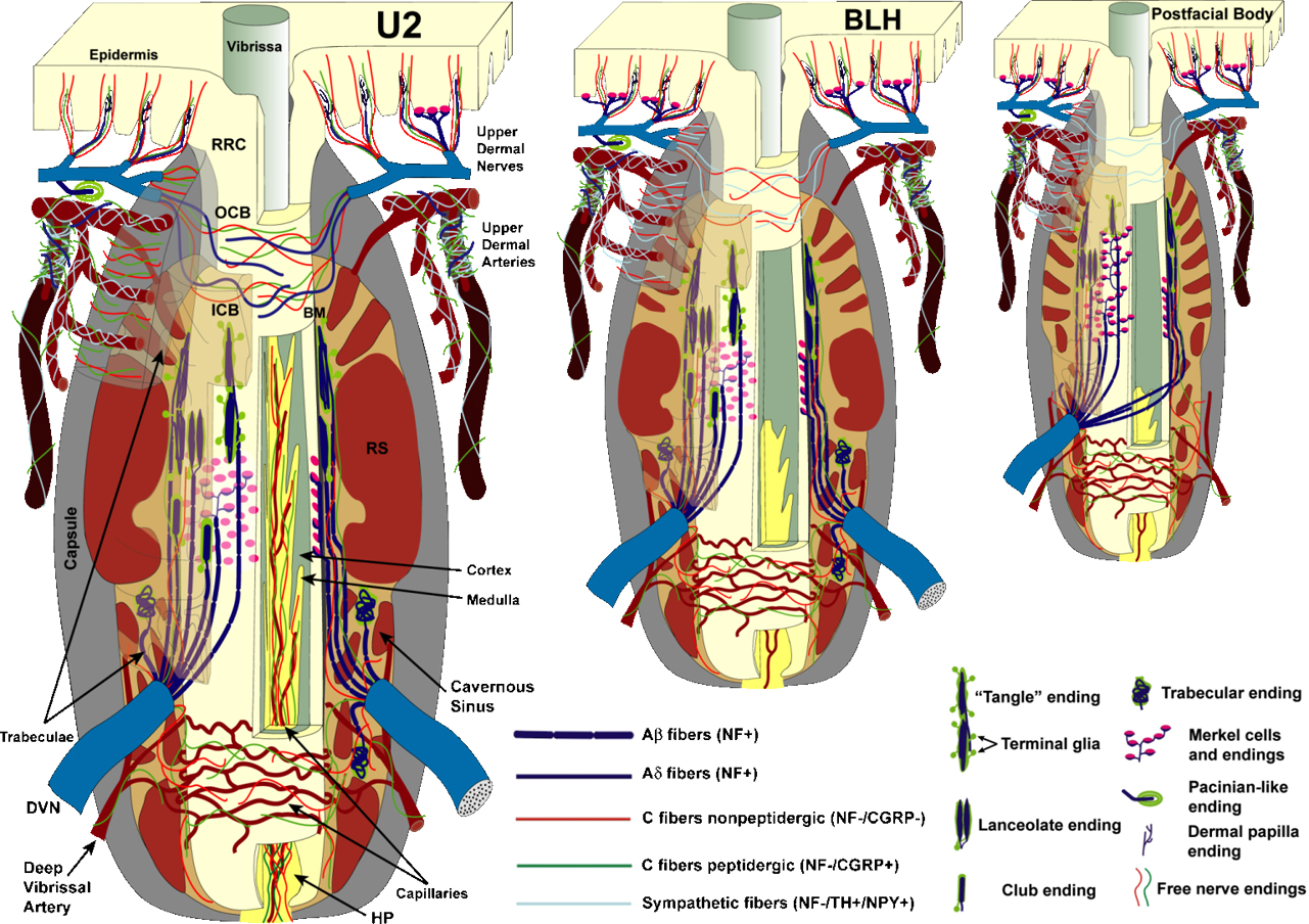
Schematic diagrams of innervation patterns for large U2 perioral vibrissae, bristle-like hairs of the oral disk, and postfacial vibrissae. From Sarko et al. (2007a).
Behavioral use of tactile hairs (vibrissae) by manatees
All sirenian hairs are vibrissae, distributed over the entire face and postcranial body, in contrast to the restricted distribution seen in other mammals. This suggests an expanded functional role for vibrissae in sirenians. We hypothesize that the perioral bristles and bristle-like hairs of the face are used most often in direct tactile contact, whereas hairs of the postcranial body serve primarily as receptors of hydrodynamic stimuli associated with movement of other animals, water currents, tidal flows and changes in topographic contours of the shallow water environment.
Our behavioral experiments utilizing two captive manatees at Mote Marine Laboratory are testing this hypothesis by comparing the sensitivity of the facial vibrissae with that of postcranial vibrissae. An underwater vibrating sphere generates vibratory stimuli of known frequency and amplitude. Subjects are trained to respond in order to indicate whether they did or did not perceive the stimulus. By varying the location of the stimuli relative to the body, and the frequency and amplitude of stimuli across many trials, we are constructing a ‘tactogram’ for the use of vibrissae by manatees. We also intend to determine the abilit
y of manatees to identify the direction from which a stimulus originates.
We hypothesize that peripheral neuroanatomical specializations associated with the vibrissae play a major role in shaping the configuration of the central somatosensory system. The proposed anatomical studies will utilize lipophilic axon tracing to investigate patterns of connections in previously defined somatosensory regions of the brainstem, thalamus, and cerebral cortex. Fresh postmortem manatee brains will be obtained from the Marine Mammal Pathobiology Laboratory in St. Petersburg, Florida.
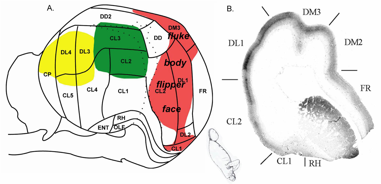
A: Putative primary somatosensory areas (red), based upon cytochrome oxidase staining patterns and cytoarchitecture. Inset shows hypothesized topography of the body map within SI. Putative AI in green, VI in yellow. B: Dense cytochrome oxidase staining in layer IV of SI in a coronal section. Courtesy of Diana Sarko.
Due to the manatee’s status as an endangered species, traditional electrophysiological methods of ascertaining the location of somatosensory regions within the CNS are not feasible. Fortunately, experimental findings in an array of species have shown that anatomical staining patterns correlate well with electrophysiological localization of sensory regions of the brainstem, thalamus and cerebral cortex. Based on a study from our lab involving manatee brain sections stained for cytochrome oxidase, acetylcholinesterase, myelin and Nissl bodies, manatees exhibit CNS somatosensory specializations including large, lobulated brainstem nuclei for processing information from oral disk vibrissae and perioral bristles (trigeminal nuclei), and from the vibrissae on the forelimb flipper and trunk (cuneate-gracile complex). A large Bischoff’s nucleus in the caudal brainstem represents input from the fluke. Bischoff’s nucleus is present in many tailed animals. In raccoons it represents the tail and projects heavily to the somatosensory thalamus. In manatees, large, subdivided ventral posterior thalamic nuclei that receive input from the brainstem somatosensory nuclei constitute a disproportionately large volume of the thalamus. The trigeminal-recipient ventral posteromedial thalamic nucleus (VPM) and the ventral posterolateral nucleus (VPL), which receives afferents associated with the fluke, body and flipper regions, are comparable in size.
Due to the manatee’s status as an endangered species, traditional electrophysiological methods of ascertaining the location of somatosensory regions within the CNS are not feasible. Fortunately, experimental findings in an array of species have shown that anatomical staining patterns correlate well with electrophysiological localization of sensory regions of the brainstem, thalamus and cerebral cortex. Based on a study from our lab involving manatee brain sections stained for cytochrome oxidase, acetylcholinesterase, myelin and Nissl bodies, manatees exhibit CNS somatosensory specializations including large, lobulated brainstem nuclei for processing information from oral disk vibrissae and perioral bristles (trigeminal nuclei), and from the vibrissae on the forelimb flipper and trunk (cuneate-gracile complex). A large Bischoff’s nucleus in the caudal brainstem represents input from the fluke. Bischoff’s nucleus is present in many tailed animals. In raccoons it represents the tail and projects heavily to the somatosensory thalamus. In manatees, large, subdivided ventral posterior thalamic nuclei that receive input from the brainstem somatosensory nuclei constitute a disproportionately large volume of the thalamus. The trigeminal-recipient ventral posteromedial thalamic nucleus (VPM) and the ventral posterolateral nucleus (VPL), which receives afferents associated with the fluke, body and flipper regions, are comparable in size.
Within cerebral cortex, the large presumptive somatosensory cortex contains multiple areas distinguished by cytoarchitecture, cytochrome oxidase staining patterns, and the presence of neuron aggregates (Rindenkerne) in layer VI that may correspond to barrels in other taxa and thus represent individual vibrissae. We also found that the putative SI of the manatee is disproportionately large (based on quantitative analysis of cytochrome oxidase-stained flattened sections), comparable to SI in other somatosensory specialists like the naked mole-rat. Our experiments using lipophilic tracers will build upon this foundation and expand our understanding of the patterns of connections among these somatosensory regions of the manatee brain.
Comparative Vascular Morphology of Head and Neck in Seals, Manatees, and Dolphins, with Emphasis on Venous Drainage of the Brain
The morphology of blood vessels supplying and draining the head of these animals is poorly understood. The greatest gaps in our knowledge involve the venous component of this circulation. These animals have been shown to possess numerous elaborate vascular adaptations which allow them to cope with the various challenges they face in the aquatic environment. As such, my goal is to elucidate the circulatory patterns in the head and neck in order to better understand how their vascular system functions as a whole, thereby allowing them to live at what might be considered the limit of physiological tolerance for an air-breathing mammal.
Fig. 1 (Carotid arteries, lateral view): Left lateral view of a volumetric (3D) reconstruction of arteries and veins in the head of a bottlenose dolphin. Blood vessels of a stranded dolphin were injected with a contrast medium and imaged through a computed tomography (CT) scanner. Imaging was performed on software donated by Dolphin Imaging & Management Solutions.
Fig. 2 (Intracranial veins, right dorsolateral view): Right dorsolateral view of a volumetric (3D) reconstruction of the dural veins draining the brain of a bottlenose dolphin. Blood vessels of a stranded dolphin were injected with a contrast medium and imaged through a computed tomography (CT) scanner. Imaging was performed on software donated by Dolphin Imaging & Management Solutions.
Movie 1 (click to see the CT imaging): Volumetric (3D) reconstruction of arteries and veins in the head of a bottlenose dolphin. Blood vessels of a stranded dolphin were injected with a contrast medium and imaged through a computed tomography (CT) scanner. Imaging was performed on software donated by Dolphin Imaging & Management Solutions.
Movie 2 (click to see the CT imaging): Volumetric (3D) reconstruction of arteries and veins in the head of a manatee. Blood vessels of the manatee were injected with a contrast medium and imaged through a computed tomography (CT) scanner. Imaging was performed on software donated by Dolphin Imaging & Management Solutions.
For more information please visit Dr. Reep’s profile which contains contact information and the Publications page.


